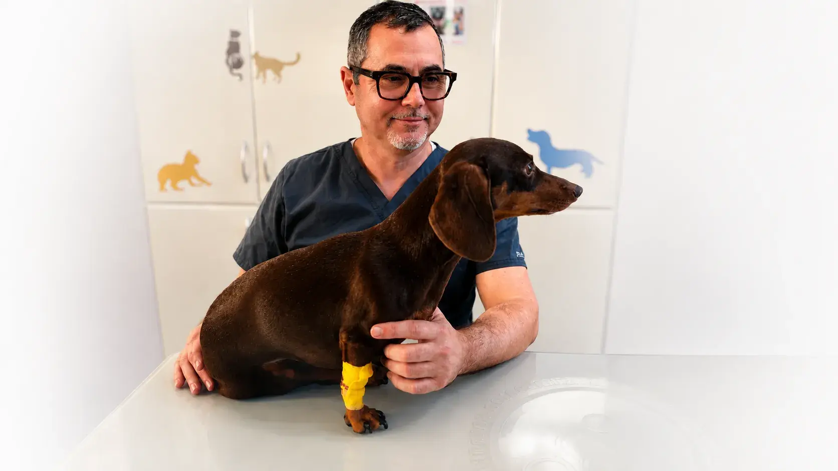
MRI, CT or X-ray — when is each one used?
Every imaging method has its place. We explain which diagnostic is right for which kind of disease.
Modern veterinary medicine uses several imaging methods. Each has its strengths and specific indications. At BENNETT we have X-ray, ultrasound, CT and MRI available.
X-ray
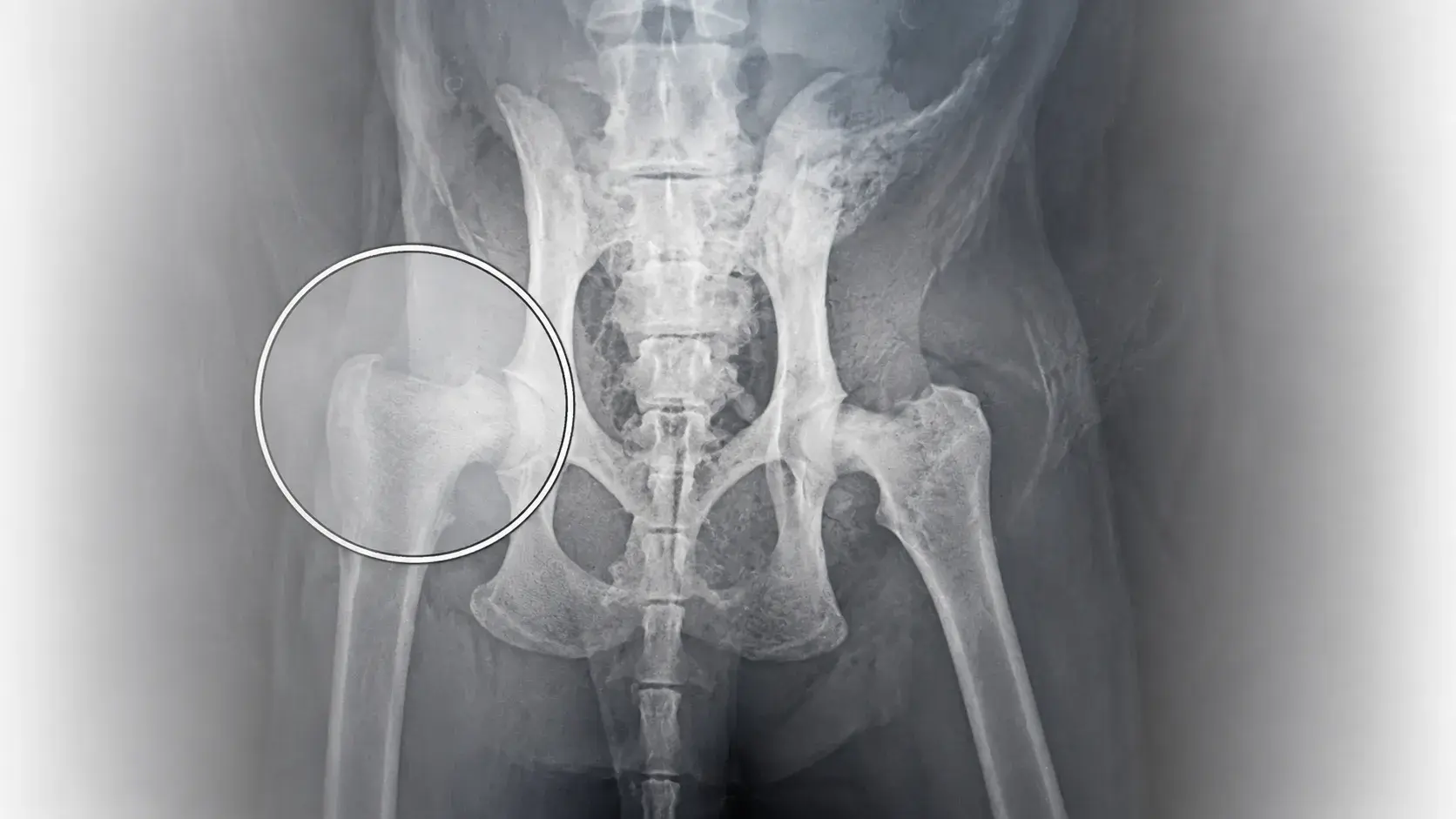
Radiography is fast, accessible and ideal for bones, joints and the chest. Suitable for suspected fractures, arthrosis, pneumonia or heart disease.
Ultrasound
Ultrasound is irreplaceable for examining the abdominal organs — kidneys, liver, spleen, uterus, bladder. It needs no anaesthesia and is safe even for pregnant females.
CT (computed tomography)
CT provides detailed 3D images of bones, blood vessels and organs. Ideal for planning orthopaedic surgery, oncological staging, or examining the chest and abdomen.
MRI (magnetic resonance imaging)
MRI is the gold standard for neurological and soft-tissue examinations — spinal cord, brain, intervertebral discs, tendons and ligaments. It requires general anaesthesia.
The right choice of imaging depends on the clinical suspicion. Our vets guide you from the first sign to the diagnosis.